iBet uBet web content aggregator. Adding the entire web to your favor.


Link to original content: http://pubmed.ncbi.nlm.nih.gov/15226428/
Six1 and Eya1 expression can reprogram adult muscle from the slow-twitch phenotype into the fast-twitch phenotype - PubMed Skip to main page content
U.S. flag

An official website of the United States government

Dot gov

The .gov means it’s official.
Federal government websites often end in .gov or .mil. Before sharing sensitive information, make sure you’re on a federal government site.

Https

The site is secure.
The https:// ensures that you are connecting to the official website and that any information you provide is encrypted and transmitted securely.

Access keys NCBI Homepage MyNCBI Homepage Main Content Main Navigation
. 2004 Jul;24(14):6253-67.
doi: 10.1128/MCB.24.14.6253-6267.2004.

Six1 and Eya1 expression can reprogram adult muscle from the slow-twitch phenotype into the fast-twitch phenotype

Affiliations

Six1 and Eya1 expression can reprogram adult muscle from the slow-twitch phenotype into the fast-twitch phenotype

Raphaelle Grifone et al. Mol Cell Biol. 2004 Jul.

Abstract

Muscle fibers show great differences in their contractile and metabolic properties. This diversity enables skeletal muscles to fulfill and adapt to different tasks. In this report, we show that the Six/Eya pathway is implicated in the establishment and maintenance of the fast-twitch skeletal muscle phenotype. We demonstrate that the MEF3/Six DNA binding element present in the aldolase A pM promoter mediates the high level of activation of this promoter in fast-twitch glycolytic (but not in slow-twitch) muscle fibers. We also show that among the Six and Eya gene products expressed in mouse skeletal muscle, Six1 and Eya1 proteins accumulate preferentially in the nuclei of fast-twitch muscles. The forced expression of Six1 and Eya1 together in the slow-twitch soleus muscle induced a fiber-type transition characterized by the replacement of myosin heavy chain I and IIA isoforms by the faster IIB and/or IIX isoforms, the activation of fast-twitch fiber-specific genes, and a switch toward glycolytic metabolism. Collectively, these data identify Six1 and Eya1 as the first transcriptional complex that is able to reprogram adult slow-twitch oxidative fibers toward a fast-twitch glycolytic phenotype.


Figures

FIG. 1.
FIG. 1.
The MEF3 element is required for specific expression of pM in adult fast-twitch skeletal muscle. Fast TA and slow Sol muscles were transfected with 3.5 μg of pM164CAT and pM125CAT plasmids as well as with 0.5 μg of pRSV-luciferase plasmid (the latter to allow normalization of transfection efficiencies). At 7 days after transfection, muscles were isolated and levels of activity of pM164CAT and pM125CAT in TA and Sol muscles (means ± standard errors of the means) were determined. Values for CAT activity are expressed relative to the level obtained in fast TA after transfection of pM164CAT (arbitrarily set as 100%). Differences in pM164CAT activity levels in fast TA muscle are represented. Each point represents the mean of six experiments.
FIG. 2.
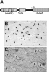
FIG. 2.
Detection of polyMEF3-nlsβgal activity in MyHCIIB fibers of transgenic animals. (A) Schematic representation of the polyMEF3-nlsβgal transgene. (B) nlsβgal activity on a transverse section of the longissimus dorsi muscle of a polyMEF3-nlsβgal transgenic adult animal. (C) Immunohistochemical analysis of MyHCIIB expression on a serial cross section. Note that only MyHCIIB positive fibers express the transgene.
FIG. 3.
FIG. 3.
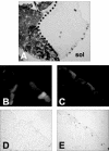
Six1 is enriched in fast-twitch muscle nuclei. (A to C) Immunohistochemistry assays of adult mouse muscle sections at the Sol-Gas level performed using polyclonal antibodies directed against Six1 (A), Six4 (B), and Six5 (C). (D) Northern blot experiment performed with 20 μg of total RNA, showing the accumulation of Six1 mRNAs (upper panel) in adult muscles together with the relative levels of 18S rRNA content (lower panel) present in the different samples analyzed. Lane 1, Gas; lane 2, extensor digitorum longus; lane 3, Sol. (E) Western blot experiment performed with 100 μg of total protein, showing the presence of Six1 (upper) and α-tubulin (below) in Gas (lane 1) and Sol (lane 2) muscles.
FIG. 4.
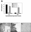
FIG. 4.
Forced expression of Six1 in adult slow Sol muscles. (A) fast TA and slow Sol muscles were transfected with 3.5 μg of pM164CAT or pM125CAT plasmids, together with 0.5 μg of pRSV-luciferase to control for transfection efficiency, and either pCMV-Six1 expression plasmid (100 ng) or empty pCR3 vector (100 ng). Values for CAT activity at 7 days after transfection of pM164CAT into Sol muscles and pM125CAT into TA or Sol muscles (means ± standard errors of the means) are expressed relative to the level obtained with 100 ng of pCR3-pM164CAT in TA muscles (arbitrarily set as 100%). Each point represents the mean of six experiments. (B) Serial section at the distal hindlimb level of untransfected pM-AP transgenic line, showing that AP activity is not detectable in Sol muscles. The inset in the right bottom corner shows a macroscopic view of the Sol, with a border of positive-testing Gas fibers. (C) Serial sections of Sol muscles transfected with 500 ng of pCMV-Six1 expression vector in pM-AP transgenic mice. At 7 days after transfection, AP activity was revealed histochemically in the Gas-Sol muscles; while most Gas fibers express the AP transgene, none of the transfected Sol fibers express AP (the position of the Sol muscle adjacent to the plantaris-gas muscle masses is demarcated by a solid line). (D) Six1-transfected fibers of the Sol show a detectable amount of Six1 protein accumulation in their nuclei, as revealed by immunochemistry with Six1 antibodies. Note that panel D is an enlargement of a Sol serial section of panel C which has been revealed with Six1 antibodies.
FIG. 5.
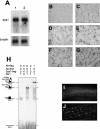
FIG. 5.
Eya1 is also enriched in the nuclei of fast-twitch muscle fibers. (A) Northern blot experiment performed with 4 μg of poly(A)+ mRNA, showing the accumulation of Eya1 mRNAs in adult mouse muscles (upper panel) together with the relative β-actin mRNA content (lower panel). Lane 1, Gas; lane 2, Sol. (B to G) Immunohistochemistry performed on adult Gas and Sol muscle sections with Eya1 (B and C, respectively), Eya2 (D and E, respectively), and Eya4 (F and G, respectively) antibodies. (H) Gel mobility shift assay performed with in vitro-translated Six1 and Eya1 proteins. Lane 1, 1 μl of in vitro-translated Six1; lane 2, 4 μl of in vitro-translated Flag-Eya1; lane 3, 1 μl of in vitro-translated Six1 plus 3 μl of in vitro-translated Eya1; lane 4, 1 μl of in vitro-translated Six1 plus 3 μl of in vitro-translated Eya1 plus 1 μl of Six1 antibodies; lane 5, 1 μl of in vitro-translated Six1 plus 3 μl of in vitro-translated Eya1 plus 1 μl of Flag antibodies. The results of fluorescent imaging of Sol muscle transfected 4 days earlier with 50 ng of Yfp-Eya1 alone (I) or 50 ng of Yfp-Eya1 in the presence of 500 ng of pCMV-Six1 expression vector (J). Magnification in panels I and J, ×200.
FIG. 6.
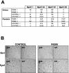
FIG. 6.
The slow to fast MyHCIIA transition observed in Sol fibers of mice treated with FK506 does not lead to Six1/Eya1 nuclear enrichment. (A) The percentages of fibers expressing different MyHC isoforms in Sol and plantaris muscles of mice treated with FK506, as well as in those of untreated controls, were determined by immunohistochemistry assays performed on hindlimb muscle cross sections at the Gas-Sol-plantaris level (n = 2 mice per group). (B) Immunohistochemical analysis of Six1 and Eya1 expression on cross-sections of FK506-treated or control Gas and Sol muscles, revealing the absence of Six1 or Eya1 nuclear enrichment in Sol fibers of FK506-treated animals.
FIG. 7.
FIG. 7.
Forced expression of Six1/Eya1 in the adult slow Sol muscle activates pM-AP and MLC3f-βgal transgenes. (A) Sol muscle cross sections obtained from pM-AP transgenic mice transfected with Six1/Eya1 expression vectors. At 2 weeks after transfection, AP activity was detected within the transfected Sol muscles of pM-AP transgenic mice. (B to E) Gfp and β-Gal activity in Sol muscles of MLC3f-nlsβgal transgenic mice transfected with a control green fluorescent protein (Gfp) expression plasmid to allow identification of transfected fibers in the presence of Six1 and Eya1 expression vectors (C and E) or of pCR3 control plasmid (B and D). In panel C, transfected Gfp-positive fibers within the Sol corresponded to fibers demonstrating nuclear β-Gal staining (E). Note that the β-Gal blue nuclei are at the periphery of the fibers, indicating that these fibers have not undergone regeneration.
FIG. 8.
FIG. 8.
Forced expression of Six1 and Eya1 in the adult slow Sol muscle drives a slow to fast transition 2 weeks after transfection. Immunohistochemical analysis at the Sol-Gas muscle level of expression with serial cross sections from pM-AP transgenic mice (B to F) or wild-type mice (G to N) transfected with Six1/Eya1 expression vectors (except in panels G and H, which show expression of MyHCI and MyHCIIX for a wild-type, not transfected, Sol section). Note that the antibody used to reveal MyHCIIX recognizes all MyHCs except MyHCIIX; thus, pure MyHCIIXs in panel H are unstained or are only weakly stained, as exemplified by those fibers marked by an asterisk in the Gas muscle image (D). (A) Schematic representation of the Gas-Sol muscle portions as presented in panels B to F; (B) AP detection; (C) MyHCIIB expression; (D) MyHCIIX expression; (E) MyHCIIA expression; (F) MyHCI expression. In panels B to F, the three long arrows indicate the same three fibers on serial cross sections, revealing that pM-AP-positive fibers also express MyHCHIIB, and perhaps MyHCIIX, but do not express MyHCIIA or MyHCI. The short arrow shows the same fiber on serial cross sections expressing MyHCIIB but not pM-AP; also note that this fiber no longer expresses either MyHCI or MyHCIIA. (G) MyHCI expression in wild-type Sol muscles at the periphery near the fibula. (H) MyHCIIX expression in Gas (left side of panel) and Sol (right side of panel) muscles of wild-type mice. (I to N) Six1/Eya1 expression vectors have been transfected into the Sol muscles together with a pDsRed expression plasmid (to visualize transfected fibers), and serial cross sections at the periphery of the muscle near the fibula are shown. (I) SERCA-1 expression, (J) DsRed detection, (K to L) MyHCIIB expression, (M) NADH-TR detection, (N) SDH detection. Note that both SDH and NADH-TR activities are downregulated in MyHCIIB-positive fibers.
FIG. 9.
FIG. 9.
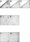
Nuclear accumulation of both Six1 and Eya1 is required to observe a slow to fast fiber type switch in Sol fibers. The results of immunohistochemical analysis of MyHCIIB expression on serial cross sections of Sol muscles transfected with 500 ng of pCMV-Six1 plus 500 ng of pCMV-Eya1 (A to C), with 500 ng of pCMV-Six1 only (D to E), or with 500 ng of pCMV-Eya1 only (F to G) are shown. MyHCIIB expression (A, D, and F) is only found within Sol fibers in which both Six1 and Eya1 nuclear accumulation are observed. Six1 nuclear expression as revealed by immunocytochemistry with Six1 antibodies (B and E). Eya1 nuclear expression as revealed by immunocytochemistry with Eya1 antibodies (C and G). Note that MyHCIIB is not detected in Sol fibers expressing only Six1 (D) or Eya1 (F). The stars in panels D and F point to the Sol fibers in which Six1 and Eya1 nuclear expression, respectively, was detected.
FIG. 10.
FIG. 10.
Slow to fast fiber type switching occurs through Six1 targets. Wild-type (A and B) or pM-AP (C to E) Sol muscles were transfected with 100 ng of pDsRed plus 500 ng of pCMV-Six1 plus 500 ng of pCMV-Eya1mutant (A and B) or with 100 ng of pDsRed plus 500 ng of pCMV-Six1-VP16 chimeric protein only (C to E). At 15 days later, transfected Sol fibers were analyzed. (A) DsRed-positive transfected fibers. (B) Absence of MyHCIIB-positive fibers in an adjacent section. (C) Immunohistochemical analysis of MyHCIIB expression. (D) AP detection. (E) MyHCemb expression on serial sections.

Similar articles

Cited by

References

    1. Abdelhak, S., V. Kalatzis, R. Heilig, S. Compain, D. Samson, C. Vincent, F. Levi-Acobas, C. Cruaud, M. Le Merrer, M. Mathieu, R. Konig, J. Vigneron, J. Weissenbach, C. Petit, and D. Weil. 1997. Clustering of mutations responsible for branchio-oto-renal (BOR) syndrome in the eyes absent homologous region (eyaHR) of EYA1. Hum. Mol. Genet. 6:2247-2255. - PubMed
    1. Anakwe, K., L. Robson, J. Hadley, P. Buxton, S. Allen, C. Hartmann, B. Harfe, T. Nohno, D. Evans, and P. Francis-West. 2003. Wnt signalling regulates myogenic differentiation in the developing avian wing. Development 130:3503-3515. - PubMed
    1. Bertrand, A., V. Ngo-Muller, D. Hentzen, J.-P. Concordet, D. Daegelen, and D. Tuil. 2003. Muscle electrotransfer as a tool for studying muscle fiber-specific and nerve-dependent activity of promoters. Am. J. Physiol. Cell Physiol. 285:C1071-C1081. - PubMed
    1. Blagden, C., P. Currie, P. Ingham, and S. Hughes. 1997. Notochord induction of zebrafish slow muscle mediated by Sonic hedgehog. Genes Dev. 11:2163-2175. - PMC - PubMed
    1. Borsani, G., A. DeGrandi, A. Ballabio, A. B. Bulfone, L. Bernard, S. Banfi, C. Gattuso, M. Mariani, M. Dixon, D. Donnai, K. Metcalfe, R. Winter, M. Robertson, R. Axton, A. Brown, V. van Heyningen, and I. M. Hanson. 1999. EYA4, a novel vertebrate gene related to Drosophila eyes absent. Hum. Mol. Genet. 8:11-23. - PubMed

Publication types

MeSH terms

LinkOut - more resources