Insulin-like growth factor 2 receptor
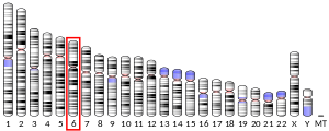
Insulin-like growth factor 2 receptor (IGF2R), also called the cation-independent mannose-6-phosphate receptor (CI-MPR) is a protein that in humans is encoded by the IGF2R gene.[5][6] IGF2R is a multifunctional protein receptor that binds insulin-like growth factor 2 (IGF2) at the cell surface and mannose-6-phosphate (M6P)-tagged proteins in the trans-Golgi network.[6]
Structure
[edit]The structure of the IGF2R is a type I transmembrane protein (that is, it has a single transmembrane domain with its C-terminus on the cytoplasmic side of lipid membranes) with a large extracellular/lumenal domain and a relatively short cytoplasmic tail.[7] The extracellular domain consists of a small region homologous to the collagen-binding domain of fibronectin and of fifteen repeats of approximately 147 amino acid residues. Each of these repeats is homologous to the 157-residue extracytoplasmic domain of the mannose 6-phosphate receptor. Binding to IGF2 is mediated through one of the repeats, while two different repeats are responsible for binding to mannose-6-phosphate. The IGF2R is approximately 300 kDa in size; it appears to exist and function as a dimer.
Function
[edit]IGF2R functions to clear IGF2 from the cell surface to attenuate signalling, and to transport lysosomal acid hydrolase precursors from the Golgi apparatus to the lysosome. After binding IGF2 at the cell surface, IGF2Rs accumulate in forming clathrin-coated vesicles and are internalized. In the lumen of the trans-Golgi network, the IGF2R binds M6P-tagged cargo.[7] The IGF2Rs (bound to their cargo) are recognized by the GGA family of clathrin adaptor proteins and accumulate in forming clathrin-coated vesicles.[8] IGF2Rs from both the cell surface and the Golgi are trafficked to the early endosome where, in the relatively low pH environment of the endosome, the IGF2Rs release their cargo. The IGF2Rs are recycled back to the Golgi by the retromer complex, again by way of interaction with GGAs and vesicles. The cargo proteins are then trafficked to the lysosome via the late endosome independently of the IGF2Rs.
Interactions
[edit]Insulin-like growth factor 2 receptor has been shown to interact with M6PRBP1.[9][10]
Evolution
[edit]The insulin-like growth factor 2 receptor function evolved from the cation-independent mannose 6-phosphate receptor and is first seen in Monotremes. The IGF-2 binding site was likely acquired fortuitously with the generation of an exonic splice site enhancer cluster in exon 34, presumably necessitated by several kilobases of repeat element insertions in the preceding intron. A six-fold affinity maturation then followed during therian evolution, coincident with the onset of imprinting and consistent with the theory of parental conflict.[11]
See also
[edit]References
[edit]- ^ a b c GRCh38: Ensembl release 89: ENSG00000197081 – Ensembl, May 2017
- ^ a b c GRCm38: Ensembl release 89: ENSMUSG00000023830 – Ensembl, May 2017
- ^ "Human PubMed Reference:". National Center for Biotechnology Information, U.S. National Library of Medicine.
- ^ "Mouse PubMed Reference:". National Center for Biotechnology Information, U.S. National Library of Medicine.
- ^ Oshima A, Nolan CM, Kyle JW, Grubb JH, Sly WS (February 1988). "The human cation-independent mannose 6-phosphate receptor. Cloning and sequence of the full-length cDNA and expression of functional receptor in COS cells". J. Biol. Chem. 263 (5): 2553–62. doi:10.1016/S0021-9258(18)69243-9. PMID 2963003.
- ^ a b Laureys G, Barton DE, Ullrich A, Francke U (October 1988). "Chromosomal mapping of the gene for the type II insulin-like growth factor receptor/cation-independent mannose 6-phosphate receptor in man and mouse". Genomics. 3 (3): 224–9. doi:10.1016/0888-7543(88)90083-3. PMID 2852162.
- ^ a b Ghosh P, Dahms NM, Kornfeld S (March 2003). "Mannose 6-phosphate receptors: new twists in the tale". Nat. Rev. Mol. Cell Biol. 4 (3): 202–12. doi:10.1038/nrm1050. PMID 12612639. S2CID 16991464.
- ^ Ghosh P, Kornfeld S (July 2004). "The GGA proteins: key players in protein sorting at the trans-Golgi network". Eur. J. Cell Biol. 83 (6): 257–62. doi:10.1078/0171-9335-00374. PMID 15511083.
- ^ Díaz E, Pfeffer SR (May 1998). "TIP47: a cargo selection device for mannose 6-phosphate receptor trafficking". Cell. 93 (3): 433–43. doi:10.1016/S0092-8674(00)81171-X. PMID 9590177. S2CID 17161071.
- ^ Orsel JG, Sincock PM, Krise JP, Pfeffer SR (August 2000). "Recognition of the 300-kDa mannose 6-phosphate receptor cytoplasmic domain by 47-kDa tail-interacting protein". Proc. Natl. Acad. Sci. U.S.A. 97 (16): 9047–51. Bibcode:2000PNAS...97.9047O. doi:10.1073/pnas.160251397. PMC 16819. PMID 10908666.
- ^ Williams C, Hoppe HJ, Rezgui D, Strickland M, Forbes BE, Grutzner F, Frago S, Ellis RZ, Wattana-Amorn P, Prince SN, Zaccheo OJ, Nolan CM, Mungall AJ, Jones EY, Crump MP, Hassan AB (November 2012). "Exon splice enhancer primes IGF2:IGF2R binding site structure and function evolution". Science. 338 (6111): 1209–1213. Bibcode:2012Sci...338.1209W. doi:10.1126/science.1228633. PMC 4658703. PMID 23197533.
Further reading
[edit]- O'Dell SD, Day IN (1998). "Insulin-like growth factor II (IGF-II)". Int. J. Biochem. Cell Biol. 30 (7): 767–71. doi:10.1016/S1357-2725(98)00048-X. PMID 9722981.
- Hawkes C, Kar S (2004). "The insulin-like growth factor-II/mannose-6-phosphate receptor: structure, distribution and function in the central nervous system". Brain Res. Brain Res. Rev. 44 (2–3): 117–40. doi:10.1016/j.brainresrev.2003.11.002. PMID 15003389. S2CID 20434586.
- Scott CD, Firth SM (2005). "The role of the M6P/IGF-II receptor in cancer: tumor suppression or garbage disposal?". Horm. Metab. Res. 36 (5): 261–71. doi:10.1055/s-2004-814477. PMID 15156403. S2CID 260166901.
- Antoniades HN, Galanopoulos T, Neville-Golden J, Maxwell M (1992). "Expression of insulin-like growth factors I and II and their receptor mRNAs in primary human astrocytomas and meningiomas; in vivo studies using in situ hybridization and immunocytochemistry". Int. J. Cancer. 50 (2): 215–22. doi:10.1002/ijc.2910500210. PMID 1370435. S2CID 22632952.
- Zhou J, Bondy C (1992). "Insulin-like growth factor-II and its binding proteins in placental development". Endocrinology. 131 (3): 1230–40. doi:10.1210/en.131.3.1230. PMID 1380437.
- Morgan DO, Edman JC, Standring DN, Fried VA, Smith MC, Roth RA, Rutter WJ (1987). "Insulin-like growth factor II receptor as a multifunctional binding protein". Nature. 329 (6137): 301–7. Bibcode:1987Natur.329..301M. doi:10.1038/329301a0. PMID 2957598. S2CID 4308631.
- Oshima A, Nolan CM, Kyle JW, Grubb JH, Sly WS (1988). "The human cation-independent mannose 6-phosphate receptor. Cloning and sequence of the full-length cDNA and expression of functional receptor in COS cells". J. Biol. Chem. 263 (5): 2553–62. doi:10.1016/S0021-9258(18)69243-9. PMID 2963003.
- De Souza AT, Hankins GR, Washington MK, Orton TC, Jirtle RL (1996). "M6P/IGF2R gene is mutated in human hepatocellular carcinomas with loss of heterozygosity". Nat. Genet. 11 (4): 447–9. doi:10.1038/ng1295-447. PMID 7493029. S2CID 21787312.
- Ilvesmäki V, Blum WF, Voutilainen R (1994). "Insulin-like growth factor binding proteins in the human adrenal gland". Mol. Cell. Endocrinol. 97 (1–2): 71–9. doi:10.1016/0303-7207(93)90212-3. PMID 7511544. S2CID 22503525.
- De Souza AT, Hankins GR, Washington MK, Fine RL, Orton TC, Jirtle RL (1995). "Frequent loss of heterozygosity on 6q at the mannose 6-phosphate/insulin-like growth factor II receptor locus in human hepatocellular tumors". Oncogene. 10 (9): 1725–9. PMID 7753549.
- Schmidt B, Kiecke-Siemsen C, Waheed A, Braulke T, von Figura K (1995). "Localization of the insulin-like growth factor II binding site to amino acids 1508-1566 in repeat 11 of the mannose 6-phosphate/insulin-like growth factor II receptor". J. Biol. Chem. 270 (25): 14975–82. doi:10.1074/jbc.270.25.14975. PMID 7797478.
- Rao PH, Murty VV, Gaidano G, Hauptschein R, Dalla-Favera R, Chaganti RS (1994). "Subregional mapping of 8 single copy loci to chromosome 6 by fluorescence in situ hybridization". Cytogenet. Cell Genet. 66 (4): 272–3. doi:10.1159/000133710. PMID 8162705.
- Ishiwata T, Bergmann U, Kornmann M, Lopez M, Beger HG, Korc M (1997). "Altered expression of insulin-like growth factor II receptor in human pancreatic cancer". Pancreas. 15 (4): 367–73. doi:10.1097/00006676-199711000-00006. PMID 9361090. S2CID 42073680.
- Tikkanen R, Peltola M, Oinonen C, Rouvinen J, Peltonen L (1998). "Several cooperating binding sites mediate the interaction of a lysosomal enzyme with phosphotransferase". EMBO J. 16 (22): 6684–93. doi:10.1093/emboj/16.22.6684. PMC 1170273. PMID 9362483.
- Nykjaer A, Christensen EI, Vorum H, Hager H, Petersen CM, Røigaard H, Min HY, Vilhardt F, Møller LB, Kornfeld S, Gliemann J (1998). "Mannose 6-Phosphate/Insulin-like Growth Factor–II Receptor Targets the Urokinase Receptor to Lysosomes via a Novel Binding Interaction". J. Cell Biol. 141 (3): 815–28. doi:10.1083/jcb.141.3.815. PMC 2132758. PMID 9566979.
- Díaz E, Pfeffer SR (1998). "TIP47: a cargo selection device for mannose 6-phosphate receptor trafficking". Cell. 93 (3): 433–43. doi:10.1016/S0092-8674(00)81171-X. PMID 9590177. S2CID 17161071.
- Wan L, Molloy SS, Thomas L, Liu G, Xiang Y, Rybak SL, Thomas G (1998). "PACS-1 defines a novel gene family of cytosolic sorting proteins required for trans-Golgi network localization". Cell. 94 (2): 205–16. doi:10.1016/S0092-8674(00)81420-8. PMID 9695949. S2CID 15027198.
- Killian JK, Jirtle RL (1999). "Genomic structure of the human M6P/IGF2 receptor". Mamm. Genome. 10 (1): 74–7. CiteSeerX 10.1.1.564.5806. doi:10.1007/s003359900947. PMID 9892739. S2CID 20181915.
- Kumar S, Hand AT, Connor JR, Dodds RA, Ryan PJ, Trill JJ, Fisher SM, Nuttall ME, Lipshutz DB, Zou C, Hwang SM, Votta BJ, James IE, Rieman DJ, Gowen M, Lee JC (1999). "Identification and cloning of a connective tissue growth factor-like cDNA from human osteoblasts encoding a novel regulator of osteoblast functions". J. Biol. Chem. 274 (24): 17123–31. doi:10.1074/jbc.274.24.17123. PMID 10358067.
External links
[edit]- Insulin-Like-Growth-Factor+II+Receptor at the U.S. National Library of Medicine Medical Subject Headings (MeSH)


